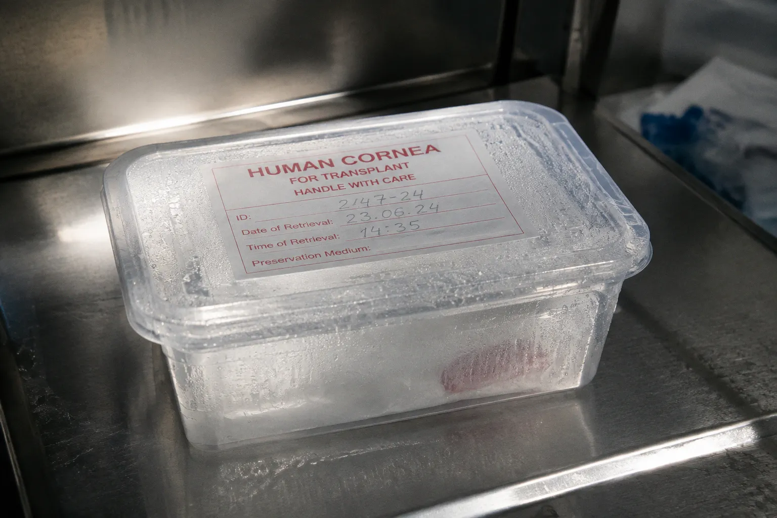
Somewhere in a clean, temperature-controlled storage medium, a cornea could still be viable — but the window is narrowing, and not because of biology alone. It is narrowing because of unclosed eyelids, because of an overhead fan left running, because of a registrar who never asked the right question at the right moment. The cornea is one of the most routinely transplanted tissues in modern medicine, and yet it is also one of the most quietly squandered. The bottleneck is not surgical skill or laboratory capacity. It is the gap between death and retrieval — and the small, mundane failures that occur inside it.
The cornea is not a relic of the deceased. It is a piece of public infrastructure, and it degrades on the same schedule as anything else left unattended.
The Biology of the Retrieval Window: Why Timing Matters for Corneal Viability
Corneal tissue is uniquely unforgiving. Unlike kidneys or livers, which require perfusion and intricate surgical choreography, the cornea is avascular — it gets its oxygen directly from tears and ambient air. That anatomical simplicity is also its vulnerability. Once the heart stops, the epithelial cells on the corneal surface begin drying out within minutes, especially if the eyelids are not closed. The clock that governs tissue viability is therefore not a clinical one in the usual sense. It is a logistical one, and it starts the moment the monitor flatlines.
Standard eye banking guidelines specify that corneal retrieval should occur as promptly as possible after death. Under typical conditions, eye banks prefer to work within a four-to-six-hour window. If the body is cooled or refrigerated — and cooled properly — that window can extend significantly, in some protocols up to twenty-four hours or beyond. These are not generous margins. They are triage thresholds. The longer the interval without adequate cooling, the lower the endothelial cell count the eye bank is likely to find at screening, and the less useful the tissue becomes for transplant.
The biochemistry of epithelial decay explains why. The corneal endothelium — a single layer of cells on the inner surface that pumps fluid out of the stroma and keeps the tissue transparent — does not regenerate. Once it is damaged, whether by hypoxia, desiccation, or osmotic shock, it cannot be repaired. The eye bank can evaluate cell density at screening, but it cannot manufacture new endothelial cells. That is why the retrieval interval matters not just for logistics but for outcome: a cornea recovered late, after prolonged exposure to warm, dry conditions, may technically pass anatomical inspection but carry a cell count too low for a reliable long-term graft.
The Cornea Preservation Time Study, a major clinical trial, demonstrated that donor corneas could maintain acceptable endothelial cell density after extended storage in modern preservation media — under the right conditions, for up to two weeks. But preservation time after recovery and time-to-retrieval after death are different clocks. The preservation clock begins when the cornea enters the storage medium; the retrieval clock begins when the heart stops. The second clock runs faster, is less forgiving, and is the one behind most of the failures eye banks report.
| Timeframe | Clinical Status | Eye Bank Decision Profile |
|---|---|---|
| 0–6 hours post-mortem | Ideal retrieval window | Tissue routinely accepted |
| 6–8 hours, body cooled/ambient-protected | Extended window, conditional | Accepted with documentation of conditions |
| Up to 24 hours, body refrigerated promptly | Preserved window | Accepted if cold chain confirmed |
| Past 24 hours, no refrigeration | Tissue compromise likely | Frequent rejection at screening |
A cornea retrieved at four hours is not the same product as one retrieved at fourteen. The donor's gift is identical; the tissue quality is not.
Immediate Post-Mortem Care: Preventing Desiccation and Epithelial Damage
Here is where the system reveals its quiet dysfunction. The retrieval window is governed less by surgical infrastructure than by a handful of bedside decisions that happen before any eye bank coordinator arrives on the scene. Four interventions, none of them exotic, determine whether tissue survives long enough to be evaluated:
- Closing the deceased's eyelids. An eye that remains open after death begins losing its epithelial surface within minutes. Overhead airflow — from a ceiling fan, a ventilator, an open window — accelerates this. A pair of closed lids is the single most important barrier between a viable cornea and a dried-out one.
- Elevating the head. Roughly thirty degrees, or simply a pillow under the head and shoulders, reduces facial edema and helps maintain the tear film that protects the epithelium during the post-mortem interval.
- Moist cotton or gauze over the closed eyes. This buys time. It does not replace refrigeration, but it slows desiccation during the interval before the body is moved.
- Switching off overhead fans and direct airflow. A simple environmental adjustment that, in practice, is almost never made without being prompted.
These are not advanced interventions. They require no specialist training, no equipment beyond what is already in the mortuary or the ward, and no special authorization. They require only that the staff on duty understand what is at stake. The uncomfortable truth is that the most expensive piece of medical equipment in this pipeline is the attention of a nurse or a junior doctor who has been told — or has not been told — that eyelids need closing in a specific way for a specific reason that has nothing to do with cosmetics.
The discrepancy is structural. Hospitals invest heavily in operating theatres, in cold-chain logistics for solid organs, in transplant coordinators on salary. They invest almost nothing in training ward staff to recognize that the deceased on bed six is, for the next few hours, a potential donor, and that their behavior in the next few minutes will determine whether an eye bank ever sees that tissue. The result is a routine of omission: fans left running, eyes left open, heads flat, consent conversations deferred to the morning shift.
A few minutes of simple bedside care — closed eyelids, elevated head, no direct airflow — can mean the difference between a viable graft and a wasted opportunity. The window does not wait for the morning shift.
Navigating Consent Hurdles: Overcoming Delays in Donor Authorization
Biology is the constraint that gets discussed in textbooks. Consent is the constraint that gets discussed in hallways — usually after the window has closed. The retrieval window is unforgiving; the consent process is often glacial. These two timelines do not align, and the mismatch is one of the central inefficiencies in donation medicine.
The legal landscape is complex and varies widely by jurisdiction. In many countries and U.S. states, the deceased's prior registration as an organ and tissue donor carries legal weight, and next-of-kin consent is not strictly required to proceed — though in practice, procurement teams are understandably reluctant to override a family's objections. In other jurisdictions, family authorization remains a mandatory legal step regardless of the deceased's wishes. The result is a patchwork: in some settings, a signed donor card is enough; in others, a grieving relative who arrives hours after the death holds effective veto power over a decision the deceased already made.
The consequence, across all of these systems, is that a family — often arriving at the hospital well after the death, sometimes not at all — becomes the gatekeeper of a time-sensitive decision. When the family cannot be reached, or cannot decide, or simply does not understand what is being asked, the clock runs out.
This is not a hypothetical failure mode. It is the dominant one. Delayed family consent is among the most commonly cited causes of missed retrieval windows in published case reviews of eye banking. The tissue was medically eligible. The deceased had registered. The eye bank was on call. And none of that mattered, because the conversation that needed to happen in the first hour happened in the sixth hour — or never happened at all.
The compliance question here is not whether to respect the family's wishes — that is non-negotiable. The question is whether the system that approaches the family is competent to do so at the right time, in the right way, with the right information. A trained requester, embedded in the hospital rather than parachuted in from a regional procurement organization, can meaningfully improve consent rates. A junior clinician reading from a script at three in the morning while the family is still in shock cannot. The procurement process is bottlenecked not by scarcity of willing donors but by scarcity of trained, well-resourced conversations.
The system asks grieving strangers to make a fast decision about a stranger's body. It should not be surprised when the answer is no.
Debunking Eligibility Myths: Who Can Actually Be a Corneal Donor
The second bottleneck is informational, and it operates upstream of the bedside. Public misunderstanding of donor eligibility criteria removes a significant fraction of medically eligible donors from the pool before the question of consent is ever raised. The myths are persistent, and they cost tissue.
The disqualifying conditions for corneal donation are narrower than most people assume. Active systemic infection — HIV, hepatitis B or C, rabies, septicemia — is an absolute contraindication, as are certain neurodegenerative diseases of unknown etiology, because of the theoretical risk of prion transmission. Outside that short list, most of the common assumptions about who cannot donate are wrong.
- Poor eyesight does not disqualify a donor. The cornea is a transparent tissue; refractive error originates in the shape of the eye, not in the cornea's cellular integrity.
- Cataract surgery does not automatically disqualify. Tissue may still be usable depending on surgical history and endothelial cell count. The eye bank makes that call.
- Wearing glasses is irrelevant to eligibility. A corneal transplant replaces the tissue itself; the donor's prescription history has no bearing on the graft.
- Most systemic cancers do not contraindicate corneal donation. The cornea is avascular and is not a typical vector for metastatic spread. Tissue from donors with a history of intraocular melanoma or certain ocular malignancies is excluded, but the far more common scenario — a donor who died of lung cancer, colon cancer, breast cancer — does not by itself disqualify the corneas.
- Advanced age is not a barrier. Most eye banks set no upper age limit. Endothelial cell density declines with age, but many donors in their seventies, eighties, and even nineties provide tissue that meets screening thresholds.
- Diabetes, in most cases, does not disqualify. Tissue is evaluated individually; diabetic donors provide usable corneas routinely.
This is not a marginal distinction. Eye banks routinely accept tissue from donors who would have been screened out by a family member relying on folk knowledge — "He had cancer," "She needed thick glasses her whole life," "His eyes were bad." Each of those filters, applied by a well-meaning relative who was never corrected by a clinician, removes usable tissue from a supply that is already structurally insufficient.
The corrective is straightforward: eligibility screening belongs to the eye bank, not to the bedside nurse, not to the funeral director, and not to the family member trying to recall what their relative said once at a dinner table. The systemic error is that screening is, in practice, distributed across all of these actors, and each of them applies a different and often incorrect standard.
| Common Belief | Medical Reality |
|---|---|
| Poor vision disqualifies | No — refractive error is irrelevant |
| Cataract surgery disqualifies | Usually no, depends on individual case |
| Cancer history disqualifies | Usually no, except certain ocular cancers |
| Old age disqualifies | No upper age limit in most programs |
| Diabetes disqualifies | No, in most cases |
| Active systemic infection | Yes — this does disqualify |
The average donor who is excluded by popular myth could have given sight to two people. The system filters them out before any specialist ever sees the file.
The Surgical Reality: How Retrieval Preserves Facial Integrity
The third and final bottleneck is psychological, and it is the one that drives family refusal rates more than any clinical or logistical concern. The persistent belief that organ and tissue donation disfigures the body is, in the case of corneal donation, simply false — and the gap between public perception and surgical reality is large enough to be measurable in missed donations.
The surgical retrieval of corneal tissue is a brief procedure. It does not involve removing the eye. The technique extracts only the cornea — the clear, dome-shaped front layer — or the corneoscleral disk, which includes a small rim of surrounding scleral tissue. The globe itself remains in the socket. The eyelids are closed afterward in the normal fashion. An open-casket viewing is not affected. The donor's facial appearance is intact.
This is not a marginal claim. In accredited eye banks that follow established protocol — whether operating under the Eye Bank Association of America's standards, the European Eye Banking Association's guidelines, or equivalent national frameworks — the surgical approach is consistent: minimal dissection, no removal of the globe, no visible alteration to the donor's face. Regional practices vary in their specific protocols and regulatory oversight, but the principle of tissue-limited retrieval that preserves facial integrity is widespread in established programs. Families who decline donation because they believe their relative will be "taken apart" or "have their eyes removed" are declining a procedure that, in anatomical terms, leaves no visible trace. They are declining a myth.
One donor can restore sight to up to two people, because each cornea is evaluated and transplanted separately. That is two grafts from one retrieval, two operations, two rehabilitations. The arithmetic is generous. The constraint is not supply. The constraint is the conversation, the bedside care, the trained requester, the closed eyelids, the stilled fan, the consent that arrives before the window closes.
The procedure leaves no mark. The system's failures do.
The cornea sits at the intersection of everything that makes modern donation medicine both extraordinary and frustrating. It is a tissue that can be retrieved quickly, stored for extended periods in modern preservation media, transplanted with mature surgical technique, and sourced from a donor pool that is overwhelmingly larger than the system currently accesses. It does not require the complex matching protocols of solid organ transplantation. It does not depend on the same cold-chain logistics or waiting-list infrastructure. What it requires — and this is the recurring finding of every performance audit in eye banking — is competent execution in the hours after death.
The retrieval window is not a mystery. It is a workflow. Closing eyelids is a workflow. Switching off a fan is a workflow. Approaching a family with trained, compassionate directness at the right moment is a workflow. Correcting eligibility myths before they reach the bedside is a workflow. The tissue is available. The recipients are waiting. The question is whether the system between them will be assembled competently enough, and quickly enough, to honor the clock that has already started.