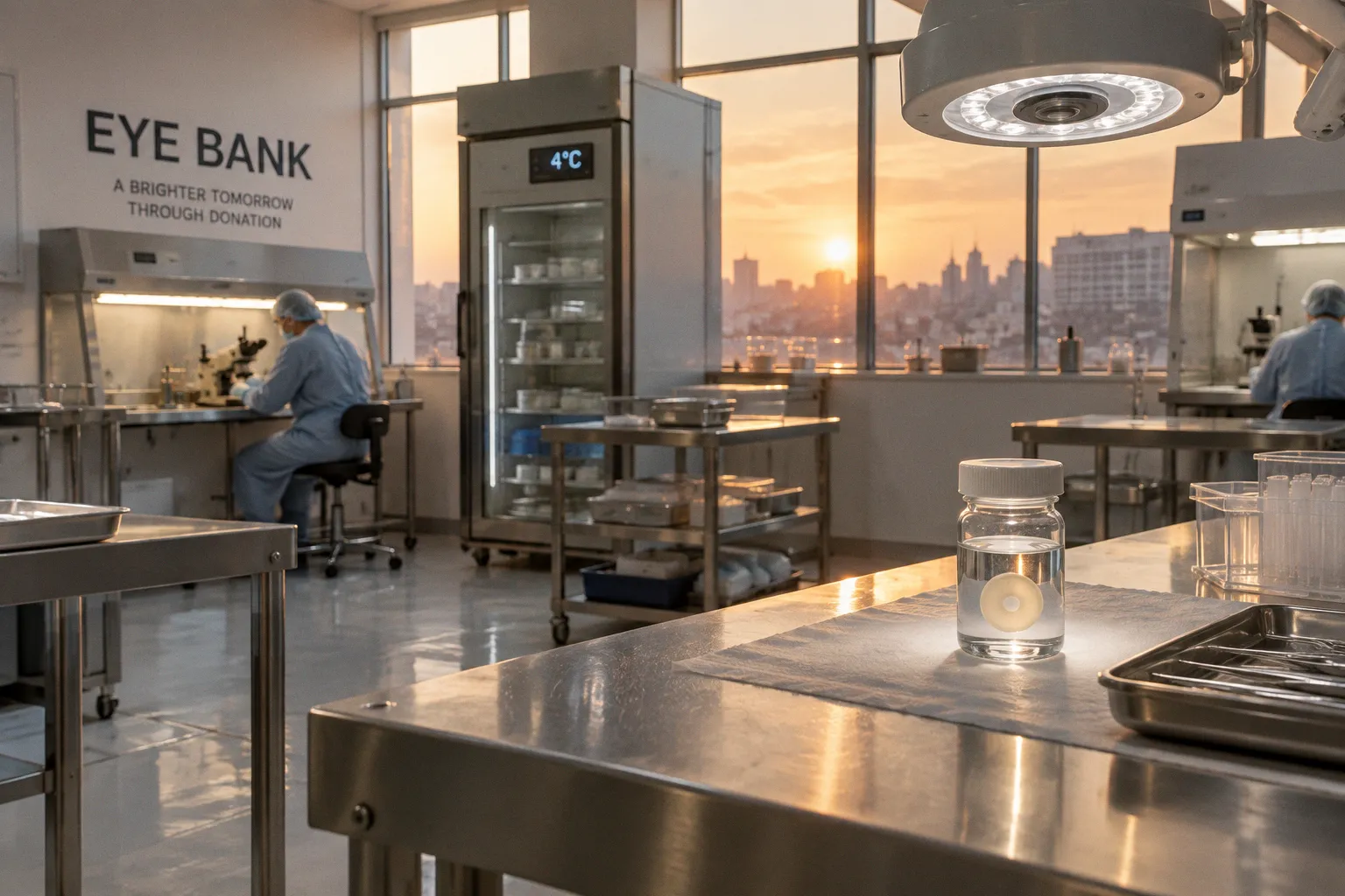
Every step is designed to answer the same urgent question: can this delicate tissue still support a clear, functioning graft when a surgeon places it into another person’s eye?
That work happens largely out of public view, in eye banking laboratories where the clock is always present. Corneal tissue must remain viable, traceable, and safe while technicians assess its quality and prepare it for procedures such as DSAEK and DMEK. The process is part laboratory medicine, part logistics, and part quiet stewardship of a gift made at the end of someone’s life.
The science of preservation: cold storage and organ culture
The cornea has no blood vessels, but it is not inert tissue. Its innermost layer contains endothelial cells that help regulate the fluid balance of the cornea and preserve its transparency. If too many of those cells are lost, the tissue may swell and become unsuitable for transplantation.
That makes preservation more complicated than simply placing a cornea in a refrigerator. The eye bank must slow cellular deterioration while protecting the tissue from contamination, maintaining the identity of the donor, and leaving enough time for evaluation and surgical planning.
Two broad preservation approaches are used in eye banking:
| Preservation method | Typical conditions | Approximate usable window | Practical implication |
|---|---|---|---|
| Hypothermic cold storage | 2°C to 8°C, typically around 4°C | Up to 14 days | Compact, established method that supports controlled transport and scheduling |
| Organ culture | 31°C to 37°C | Up to 35 days | Provides a longer preservation window but requires a different laboratory workflow and monitoring system |
Cold storage is familiar because it slows the metabolism of the corneal tissue. In media such as Optisol-GS, the tissue is kept at low temperature and monitored within a defined period. The commonly cited temperature range is 2°C to 8°C, with approximately 4°C often used as the working point. This does not turn the cornea into a shelf-stable product. The preservation window remains limited, and the tissue’s condition must still be evaluated before release.
Organ culture takes the opposite temperature approach. Rather than cooling the cornea, laboratories maintain it at approximately 31°C to 37°C in a nutrient medium. Under those conditions, donor tissue can remain viable for up to 35 days, creating more time for testing, preparation, and coordination with a transplant service.
The longer window can be especially meaningful when tissue must travel between regions or when a transplant schedule changes. But additional time does not remove the need for vigilance. Warmer culture requires a laboratory system capable of maintaining the correct environment and identifying contamination or declining tissue quality before the graft reaches a patient.
Preservation is not storage in the ordinary sense. It is a timed intervention that keeps living tissue within the narrow conditions where surgery remains possible.
Why the preservation medium matters
The fluid surrounding a donor cornea is part of the preservation strategy. It supports the tissue during transport and helps limit the cellular stress created by removal from the donor body. The medium must be handled under controlled conditions, and the container must remain linked to the donor record throughout the process.
For corneal tissue preserved in Optisol-GS, the supplied research identifies a critical endothelial cell density threshold above 2,200 cells per square millimeter. That figure is not a substitute for the full release assessment, but it illustrates why the endothelial layer receives so much attention. A cornea may look clear to the naked eye and still require detailed microscopic evaluation before it can be considered for transplantation.
In practice, the laboratory is balancing several questions at once:
- Are the endothelial cells sufficiently numerous and healthy?
- Has the tissue remained within the validated temperature and time limits?
- Is the donor record complete and correctly matched to the specimen?
- Are there signs of contamination, damage, infection, or unsuitable anatomy?
- Can the tissue be prepared for the operation requested by the surgeon?
The answers are recorded rather than assumed. A donor cornea is not accepted simply because it is available. It is released only when the available evidence supports its use.
Eye bank specular microscopy: seeing what the eye cannot
The most important details of a cornea are not visible from the outside. This is where eye bank specular microscopy becomes central to the evaluation process.
Specular microscopy allows technicians and eye bank specialists to examine the endothelial layer at high magnification. The system captures reflected light from the cell surface and produces an image that can be analyzed for cell density, cell shape, and the overall organization of the endothelial mosaic.
The endothelial cells are often described as a mosaic because they form a tightly packed layer across the back surface of the cornea. Their arrangement is not merely an aesthetic detail. It gives the laboratory information about how much functional reserve the tissue may have and whether it is likely to maintain corneal clarity after transplantation.
Donor cornea endothelial cell density
Endothelial cell density, or ECD, is usually expressed as the number of cells per square millimeter. A higher count generally indicates a stronger reserve, but the number must be interpreted alongside the image quality, cell uniformity, tissue history, and intended surgical use.
A single density figure cannot tell the entire story. The technician also looks at whether the cells are reasonably regular in shape and distribution. Variation in cell size and shape can indicate stress or reduced physiological reserve. The quality of the scan matters as well: folds, poor focus, tissue curvature, or artifacts can make an image difficult to interpret.
For corneas intended for endothelial keratoplasty, this examination becomes especially important because the transplanted tissue includes the endothelial layer that must function inside the recipient’s eye. DSAEK and DMEK are not simply procedures that replace a transparent window. They depend on the transplanted endothelial cells attaching, spreading, and regulating fluid in the recipient cornea.
A laboratory may therefore consider:
1. Cell density — whether the number of endothelial cells meets the relevant release threshold for the tissue and procedure.
2. Cell appearance — whether the cells show acceptable uniformity and organization.
3. Tissue clarity — whether the cornea can be examined and prepared without evidence of unacceptable damage.
4. Storage history — how long the tissue has been preserved and under which method.
5. Surgical purpose — whether the graft is intended for full-thickness transplantation, DSAEK, DMEK, or another form of corneal repair.
The result is a decision based on a group of findings rather than one attractive image or one isolated number.
Why microscopic evaluation remains a human task
Modern imaging systems produce measurements, but the laboratory still needs trained people to recognize limitations in the data. A low-quality scan can be misleading. Tissue folds may obscure cells. Preparation may alter the surface being measured. The technician must distinguish a genuine concern from an artifact created during imaging or handling.
This is one reason accreditation and documented experience matter. EBAA accreditation standards require an organization seeking initial accreditation for particular eye bank functions to document the handling of at least 25 surgical corneas for each function sought. The number does not transform the work into a mechanical routine, but it demonstrates that the laboratory has built a meaningful operational foundation before presenting itself as proficient in those functions.
The same principle applies to continuing oversight. Active member eye banks accredited through the Eye Bank Association of America undergo site inspections and reaccreditation at least every three years. For patients and surgical teams, that interval represents more than a line in a policy document. It is part of the groundwork that supports confidence in the tissue supply.
Precision engineering: preparing grafts for DSAEK and DMEK
Once a cornea has been evaluated and assigned for transplantation, the laboratory may prepare it for the technique planned by the surgeon. The preparation is delicate because the graft must be thin, accurately shaped, and handled without unnecessary stress to the endothelial cells.
For DSAEK, a microkeratome may be used to create a posterior lamellar graft. Standard DSAEK preparation targets a tissue thickness of approximately 90 to 120 micrometers. Ultrathin DSAEK aims for a thinner graft, generally around 40 to 90 micrometers.
These dimensions are not cosmetic refinements. Graft thickness affects how the transplanted tissue sits within the recipient cornea and can influence the visual recovery pathway. A thinner graft may reduce the amount of donor tissue inserted, while still providing the endothelial cells needed to restore corneal fluid regulation.
DSAEK and DMEK are not interchangeable preparation jobs
DSAEK includes a layer of donor stroma along with the donor Descemet membrane and endothelium. DMEK uses a much thinner graft consisting primarily of Descemet membrane and the endothelial layer. The difference changes how the tissue is cut, peeled, rolled, transported within the eye, and positioned during surgery.
For DSAEK, a microkeratome can create a controlled stromal plane. Some eye banks also prepare tissue with approaches designed to meet ultrathin thickness targets. The process requires consistency because an uneven or damaged graft can create additional difficulty for the surgeon.
DMEK preparation is more tissue-sparing in one sense and more technically demanding in another. The graft is extremely thin and can naturally scroll or roll, making orientation and handling challenging. The eye bank may provide tissue that has already been peeled and prepared, reducing some of the surgical preparation burden. But no preparation method eliminates the possibility of cell loss during handling, transport, unfolding, or implantation.
A useful way to understand the laboratory’s role is to follow the graft through its practical stages:
- Selection: the tissue is matched to the intended surgical use based on its quality and endothelial assessment.
- Preparation: the graft is cut, peeled, or shaped according to the planned procedure.
- Inspection: the tissue is checked again for visible damage, correct configuration, and traceability.
- Packaging: the graft is placed in a controlled container with its donor and tissue records.
- Release and transport: it moves to the surgical facility within the validated conditions and time limits.
- Operating-room verification: the receiving team confirms the identity and preparation before the graft is used.
At every stage, the tissue is vulnerable to mechanical stress. The cornea can be folded, touched, cut, lifted, or exposed to changes in temperature. The laboratory’s discipline lies in reducing those stresses while keeping the workflow fast enough to meet the surgical schedule.
What laser preparation changes
Laser-assisted tissue preparation can offer another route to creating a lamellar graft, depending on the eye bank’s equipment, protocols, and relationship with the surgical service. A laser may be used to create a precise plane or profile in the tissue, but precision in cutting does not make the biological material predictable in every other respect.
The graft still has to be evaluated for endothelial health. It still has to be protected during transfer. It still has to arrive with a complete record and within the conditions required for its preservation method.
That distinction matters in conversations about technology. A microkeratome or laser can improve the consistency of a physical preparation step, but it does not replace donor screening, microscopy, temperature control, or experienced handling.
Donor eligibility and the safety chain
Corneal transplantation depends on public generosity, but donation is governed by medical screening rather than goodwill alone. The eye bank reviews the donor’s medical history, examines the tissue, and assesses risks that could make the cornea unsuitable.
Localized ocular infection is one of the important exclusion areas under EBAA standards. This includes bacterial, viral, fungal, protozoal, and parasitic infections involving the eye, as well as a history of ocular herpes. These criteria exist because the transplant pathway must protect the recipient from avoidable infectious risk while also preserving confidence in the eye banking system.
The screening process is not designed to make a moral judgment about a donor. It is a clinical safeguard. A person may be willing to donate and still have tissue that cannot be released for transplantation. That decision can be difficult for families and donation coordinators, particularly when every potential graft feels precious. Yet safety standards must remain consistent precisely because the recipient may be medically vulnerable and the graft will be placed directly into the eye.
The safety chain includes more than donor eligibility. It also includes:
- Correct identification from recovery through transplantation.
- Documentation of the tissue’s storage method, temperature, and timing.
- Review of ocular history and relevant infection risks.
- Microscopic assessment of the endothelium.
- Inspection for damage or unsuitable anatomy.
- Controlled preparation for the planned procedure.
- Accreditation, inspection, and quality management within the eye bank.
This is why eye banking rarely fits the public image of a simple donation handoff. It is a network of coordinated interventions, each one narrowing uncertainty before the graft reaches the operating room.
The donor gives the tissue, but a whole neighborhood of skilled work is required before that gift can become a functioning transplant.
From a donor cornea to a surgical graft
The history of corneal transplantation shows how long this field has been building its methods. In 1905, Dr. Edward Zirm performed the first successful full-thickness penetrating corneal transplant. In 1937, Vladimir Filatov demonstrated cadaveric corneal collection after death, helping establish the principle that postmortem donation could support transplantation. Dr. R. Townley Paton established the first eye bank in 1944, and the Eye Bank Association of America was founded in 1961.
Preservation methods continued to evolve. McCarey-Kaufman medium was introduced in the 1970s, followed by the transition to Optisol-GS for hypothermic corneal preservation in the 1980s. In 2006, Mark Gorovoy introduced DSAEK with microkeratome cutting, after which eye banks increasingly became involved in preparing grafts for endothelial keratoplasty.
These milestones are not distant museum pieces. They explain why a modern eye bank contains several kinds of expertise at once: recovery coordination, sterile processing, microscopy, tissue preparation, storage science, documentation, and communication with surgical teams.
The laboratory has to work at two speeds. One is deliberate: the slow, careful review of donor records and cell images. The other is immediate: a graft may be needed for a patient whose vision is deteriorating, whose cornea is painful, or whose prior transplant is failing. A delay can affect a surgeon’s schedule and a patient’s recovery, but rushing can compromise the very tissue that makes the operation possible.
That tension is handled through protocols rather than improvisation. A strong eye bank builds resilience into the workflow so that a single unexpected event—an unsuitable tissue finding, a transport delay, a change in the surgical plan—does not bring the entire process to a stop.
The future of corneal tissue processing in eye banks
The direction of corneal tissue processing is toward greater specialization. Eye banks increasingly support surgeons not only by supplying whole donor corneas, but also by preparing grafts tailored to particular endothelial procedures.
Precut DSAEK tissue is one example. Standard grafts may target 90 to 120 micrometers, while ultrathin preparations target approximately 40 to 90 micrometers. For surgical teams, receiving a prepared graft can reduce the amount of tissue manipulation required in the operating room. For patients, the benefit may appear simply as a more coordinated surgical pathway, even though the underlying preparation began days earlier in a laboratory.
DMEK has pushed the field toward even more refined handling. The graft contains very little supporting tissue, so the eye bank’s preparation and packaging methods must account for rolling, orientation, cell protection, and ease of deployment. The technical challenge is not only to make the graft thin, but to make it usable.
At the same time, the central measures remain familiar. The tissue must be safe. The endothelial cells must be viable. The donor record must be complete. The graft must remain within its validated storage window. A new instrument can change the preparation step, but it cannot remove the need for careful judgment.
For communities, this creates a direct connection between donation and clinical infrastructure. A person who registers as an eye donor may never know the technicians, coordinators, couriers, and surgeons who handle the tissue. They may never see the microscopy image or the label on the storage container. But those unseen steps are where generosity is converted into a form that medicine can safely use.
Corneal tissue processing in eye banks is therefore both highly technical and deeply human. The microscopes, storage media, temperature controls, and cutting systems matter because a recipient is waiting on the other side of them. Behind every prepared graft is a chain of people working through the same demanding shift: protect the tissue, preserve its potential, and deliver it with enough care that sight may begin again.