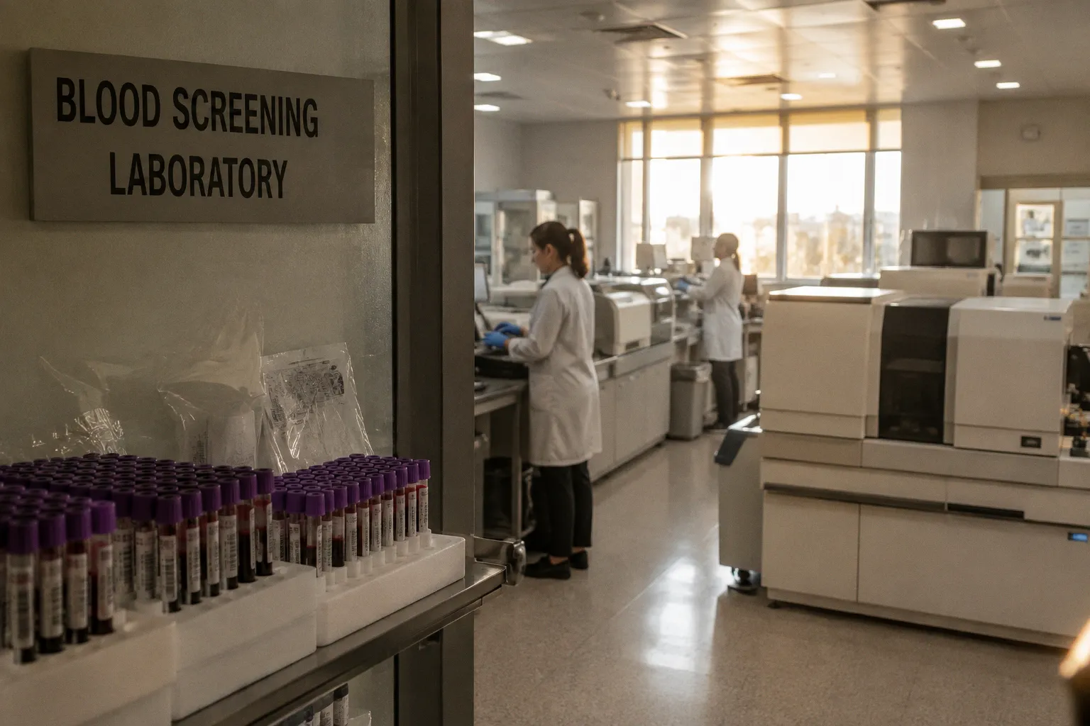
The bags come back to the regional blood center looking ordinary — a half-liter of dark red resting on a shaker — but what happens next is anything but ordinary. Before that blood touches a single patient, it travels through a layered gauntlet of laboratory work, regulatory scrutiny, and detective-level analysis designed to keep the supply safer than it has ever been in the history of transfusion medicine. The donated blood infectious disease screening process is not one test, not two. It is a chain of overlapping interventions, each one closing a gap the one before it cannot fully seal.
Understanding how that chain works matters, because every patient who receives a transfusion is trusting a process they will never see. The donor sees the arm stick. The recipient sees the bag. The middle — the cold-room logistics, the centrifuge schedule, the throughput of polymerase chain reaction machines, the lot-release paperwork — is where the actual resilience of the blood supply gets built, day after day.
Every unit of donated blood is held until it clears several independent screens. A reactive result on any one of them triggers a quiet withdrawal of that unit, long before it can be matched to a patient.
The Multi-Layered Defense: From Donor History to Laboratory Analysis
The screening process begins before a needle ever touches a vein. In a small booth near the registration table, a phlebotomist walks the donor through a lengthy health questionnaire — exposures, travel, recent illness, medications, sexual history relevant to transfusion risk. This is the first layer of defense, and for many years it carried most of the weight. In modern practice, the questionnaire is a deferral tool, not a diagnostic one. A donor who has spent three months in a malaria-endemic region is deferred for a defined period. A donor who has taken certain medications waits. A donor whose questionnaire raises a red flag never reaches the donation chair that day.
A mini-physical follows: pulse, temperature, hemoglobin check via finger-stick. The hemoglobin gate alone defers a meaningful number of would-be donors each year, particularly among young women whose iron stores run borderline. None of these steps detect infection directly. They reduce the probability that an infectious donor enters the pipeline — a probabilistic filter, not a diagnostic one.
Once the unit is collected, it enters a laboratory workflow organized around two complementary technologies. The first is serological testing — assays that look for antibodies or antigens in the donor's plasma, evidence that the immune system has responded to a pathogen or that viral proteins themselves are circulating. The second is Nucleic Acid Testing, often shortened to NAT, which looks for the genetic material of the virus directly, sidestepping the body's slower antibody response.
Neither technology alone is enough. Serological assays are highly specific and inexpensive to run in batches, but they leave a diagnostic blind spot during the early days of an infection, before the immune system has caught up. NAT closes some of that gap, but at greater per-unit cost and laboratory complexity. Run together, the two approaches answer different questions about the same unit of blood, and that redundancy is precisely the point.
Nucleic Acid Testing: Closing the Diagnostic Window Period
The diagnostic window period is the stretch of time after a donor has been infected but before any blood-based test can reliably pick up the infection. During this window, a donor feels fine, has no reason to defer, and could donate blood that passes older screening standards while quietly carrying a viral load.
Nucleic Acid Testing has changed the geography of that window. Instead of looking for what the immune system produces, NAT looks for what the virus produces — RNA or DNA sequences unique to a given pathogen. By amplifying tiny fragments of viral genetic material through polymerase chain reaction or transcription-mediated amplification, NAT can detect infections days, and sometimes weeks, earlier than serology alone.
In the United States, the American Red Cross applies a triplex NAT assay that pools donor samples into mini-pools of sixteen donations, screening each pool simultaneously for Hepatitis B virus DNA, HIV RNA, and Hepatitis C virus RNA. Pooling keeps the workflow manageable at industrial scale: a single reactive pool triggers individual retesting of every donation in that group. The FDA licensed this triplex assay in June 2009, and it has since become a backbone of routine donor screening across the national supply.
The mini-pool approach is a deliberate tradeoff. Pooling dilutes each individual donation into a larger sample, which means the analytical sensitivity per donor is lower than it would be in an individual-donation test. For most infections — most of the time — that diluted sensitivity still catches positives well within the window. For rare cases with very low viral loads, it can miss. That is why some centers, particularly during seasonal West Nile virus outbreaks or in regions with elevated risk, shift to individual-donation NAT, testing every unit on its own rather than in a pool. The per-unit cost rises sharply, but the detection threshold drops to its lowest possible level.
NAT does not make window periods vanish — it shrinks them. After its introduction for Hepatitis B, the undetected window shrank by roughly twelve days, leaving a residual window of about two to three weeks.
That residual window is small, but it is not theoretical. It is the reason every layer of screening still matters, and the reason blood banks keep investing in newer assay formats rather than relying on pooling alone.
Mandatory Pathogen Panels and Regulatory Compliance
The U.S. Food and Drug Administration requires every donation to be tested for a defined panel of infectious agents before any component can leave the blood center. The core list is precise and non-negotiable. Each donation must be screened for HIV-1 and HIV-2, Hepatitis B virus, Hepatitis C virus, Human T-Lymphotropic Virus types I and II, the bacterium that causes syphilis, West Nile virus, and Trypanosoma cruzi, the parasite responsible for Chagas disease.
The table below sketches how those mandatory screens relate to the pathogen groups and the laboratory technology most often used to detect them.
| Pathogen | Disease caused | Primary screening approach | Why one assay alone is not enough |
|---|---|---|---|
| HIV-1 / HIV-2 | AIDS | Immunoassay plus NAT for HIV RNA | Early infection predates detectable antibodies |
| Hepatitis B virus (HBV) | Hepatitis B | HBsAg, anti-HBc, plus NAT for HBV DNA | Pre-seroconversion window spans weeks |
| Hepatitis C virus (HCV) | Hepatitis C | Immunoassay plus NAT for HCV RNA | Antibody response is delayed after acute infection |
| HTLV-I / II | T-cell leukemia, myelopathy | Immunoassay | Considered acceptable as antibody-only screen |
| Treponema pallidum | Syphilis | Serology (reagin or treponemal assay) | Refrigerated storage typically limits transmission risk |
| West Nile virus (WNV) | Neuroinvasive arboviral disease | NAT-based donor screening | Viral load is detectable before antibody response; screening relies on NAT rather than serology |
| Trypanosoma cruzi | Chagas disease | Immunoassay, often once per donor | Endemic exposure history outside U.S. travel patterns |
West Nile virus deserves a closer look in that table. Unlike the other agents on the panel, WNV has no licensed serological screening test routinely used on blood donors in the United States. Surveillance relies entirely on NAT, either through mini-pooled or individual-donation testing depending on seasonal activity and regional case counts. When mosquito season drives up infection rates in a given area, blood centers in affected regions typically escalate from mini-pool to individual-donation NAT within days — a dynamic response that reflects the virus's unpredictable geography.
Beyond the core panel, additional targeted screening kicks in based on geography and recipient risk. Donors in regions endemic for Babesia species, tick-borne parasites that can infect stored red cells, are subjected to additional NAT screening. Recipients at heightened risk for complications from cytomegalovirus — particularly immunocompromised patients and low-birthweight neonates — receive CMV antibody-tested components. Some centers also screen for Trypanosoma cruzi using a one-time-per-donor model, since chronic infection persists for life and a single confirmed-negative result is sufficient to clear future donations from the same individual.
The regulatory backbone for all of this sits in the U.S. Code of Federal Regulations. Under 21 CFR 610.40(a), no blood component may be released for transfusion unless all required infectious disease tests are confirmed nonreactive. That single sentence is the operational heartbeat of every blood bank's release desk. Every morning, the supervisor checks that the donor's panel results are filed, that no test is pending, and that no reactive flag has been overridden before a unit proceeds to inventory.
The Persistent Challenge of Bacterial Contamination in Platelets
Viruses get most of the attention, but the most common infectious risk in modern transfusion medicine is bacterial, and it concentrates in one product above all others: platelets.
Unlike red cells and plasma, which are stored cold or frozen, platelets are kept at room temperature — typically 20 to 24 degrees Celsius, with gentle agitation — to preserve their clotting function. That storage condition also supports bacterial growth if any skin contaminant was introduced at the phlebotomy site. A single colony of Staphylococcus or Streptococcus introduced during needle insertion can multiply to clinically dangerous levels within 24 to 48 hours of collection.
Despite improved skin disinfection protocols, diversion pouches that discard the first few milliliters of blood, and routine culture-based screening, bacterial contamination of platelet products still occurs in roughly 1 in 2,000 to 2,500 transfusions — a stubbornly persistent risk compared with the vanishingly small per-unit residual risk of viral transmission. The contrast is worth pausing on: viral screening has driven residual risk to levels measured in millions, but bacterial contamination in platelets remains stubbornly in the low thousands.
Reducing that number has become a frontline priority. Many centers now use secondary culture steps drawn from the platelet bag 24 to 36 hours after collection, allowing any introduced organism time to replicate to detectable levels before the culture is read. Pathogen-reduction technologies — systems that use ultraviolet light combined with a photosensitizing agent to inactivate a broad spectrum of bacteria, viruses, and parasites — offer another layer and are increasingly adopted, particularly for apheresis platelets. Rapid bacterial detection assays performed on the day of transfusion add a final check at the point of issue. Each intervention adds time, cost, and labor to the workflow. Each one also adds a layer of protection for the patient who will receive the bag on day five or day seven of its shelf life. The fact that platelets are the most fragile link in the safety chain is exactly why so much of the daily craftsmanship in a modern blood bank is devoted to handling them.
Residual Risk and the Limits of Modern Screening Technology
No screening process is, or can be, perfect. The diagnostic window persists, though it grows shorter with each generation of assay. Some pathogens, particularly those with long, silent incubation periods or those not yet recognized as transfusion-transmissible, can in principle slip past current assays. The honest framing is this: donated blood is screened for the pathogens that matter most in the donor population served, using the best tools available, within an economic ceiling that allows the system to function at industrial scale.
That honest framing is part of why blood establishments treat every donation as potentially suspicious until proven otherwise. The American Red Cross and similar national systems do not just run tests; they run a layered operational culture around them. Each unit is linked to its donor by a unique identifier. When repeat and supplemental testing confirms a true-positive result, a lookback investigation is initiated to identify and notify hospitals that received prior components from the same donor — a process designed to catch any transfusion that occurred before the infection was detectable. Donors who return with a confirmed positive are entered into a national deferral registry so that future donations from the same person are blocked automatically, without relying on the donor to self-report.
The system's strength lies in the way these layers interact. A donor questionnaire catches travel and medication history. Serology catches chronic infections with mature immune responses. NAT catches early-stage infections the immune system has not yet flagged. Culture and pathogen reduction catch the bacteria that none of the above address. And the deferral registry ensures that a donor who tests positive once cannot re-enter the supply under a different name or at a different drive.
The promise of the modern blood supply is not a guarantee of zero risk. It is a system that narrows the unknown, holds every unit until it is cleared, and reacts instantly when a confirmed signal appears.
The work behind that promise is mostly invisible — performed in windowless rooms by technologists who never meet the patient on the receiving end of the line they just released. But for the regulars at the morning blood drives, the volunteers running the donor table, the recruiters working the neighborhood, and the nurses walking the bag down to the ward, this layered architecture is the quiet foundation of what they do. It is the reason a routine surgery, a complicated delivery, a chemotherapy cycle, or a trauma resuscitation can proceed without the additional fear of a transfusion-transmitted infection. The screening process will never be finished work. It is the kind of intervention that has to be rebuilt, recalibrated, and patiently defended, one unit at a time, every day.