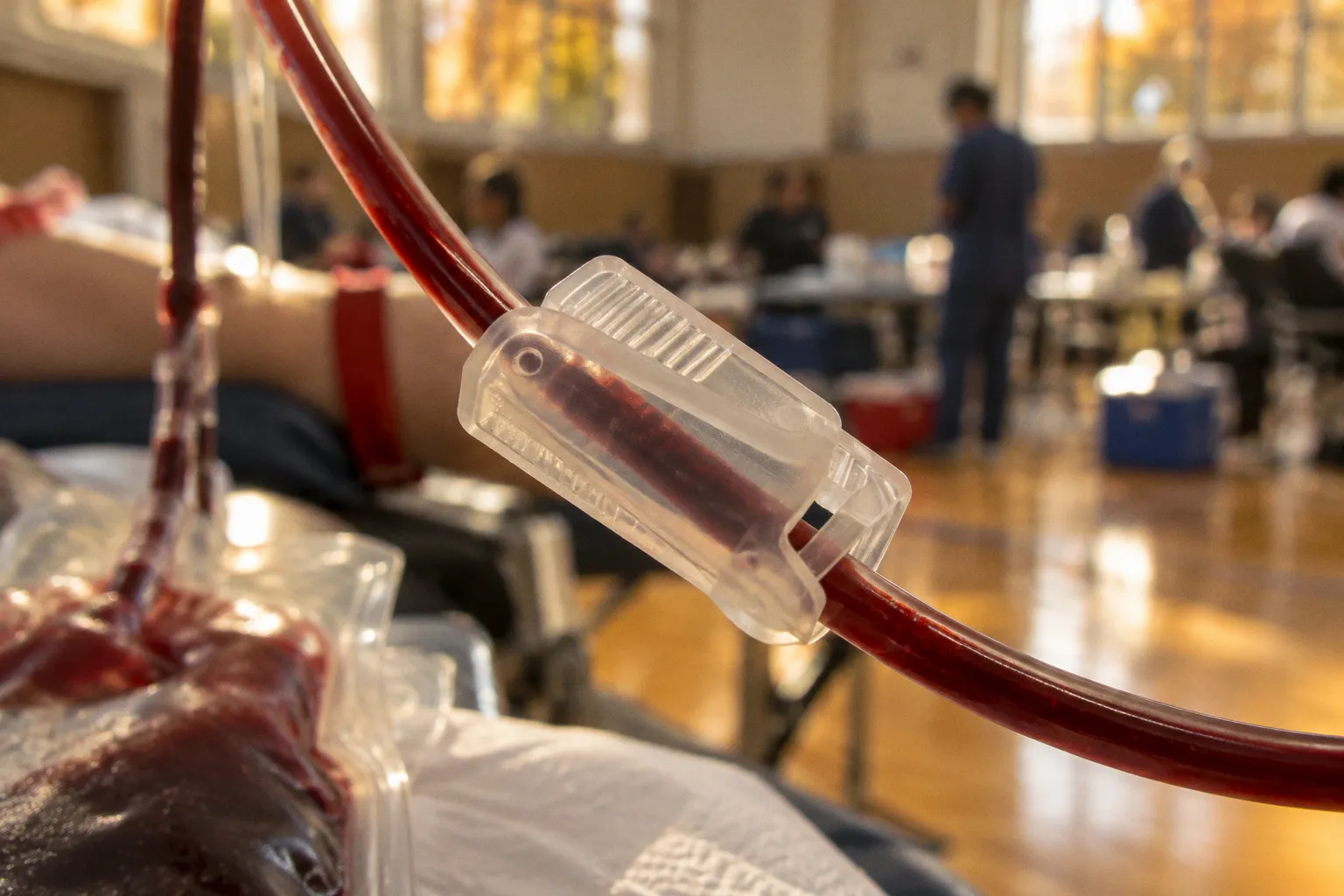
The public-facing version of blood donation tends to stop at the collection chair. The operational reality begins afterward, when a blood bank has to turn one perishable donation into components without confusing speed with control.
The blood component separation process in clinical settings is therefore less a matter of simply spinning a bag in a centrifuge than of managing density, force, temperature, timing and traceability at the same time. A small discrepancy in the protocol can affect what remains in each layer. An apparently minor documentation or storage failure can turn a usable component into regulated waste.
The central contradiction is familiar across medical logistics: blood is donated as a gesture of solidarity, but it is managed as a tightly controlled biological commodity. The system has to preserve both facts.
The physics of centrifugation: RCF is the number that matters
Whole blood contains components with different specific gravities. When a collected unit is placed in a centrifuge, controlled centrifugal force encourages those components to separate according to density. The resulting arrangement is broadly predictable:
- plasma occupies the least dense, upper portion;
- platelets are found below the plasma layer;
- leucocytes collect in the buffy coat;
- packed red blood cells form the densest lower layer.
That description sounds orderly because, under controlled conditions, it is. But blood bank laboratory workflows are not governed by visual appearance alone. The separation must be reproducible, and reproducibility depends on the force applied to the blood—not merely on how fast a rotor appears to turn.
This is where the persistent use of revolutions per minute, or RPM, becomes an administrative shortcut with technical consequences. RPM describes rotational speed. It does not describe the actual centrifugal force experienced by the sample unless the rotor radius is also known. Two centrifuges operating at the same RPM can subject blood units to different forces if their rotors have different dimensions.
The relevant metric is relative centrifugal force, or RCF, usually expressed as × g. A commonly used relationship is:
RCF = 1.118 × 10⁻⁵ × rotor radius in centimetres × RPM²
The formula is not decorative laboratory mathematics. It explains why a processing instruction that lists only RPM may be incomplete. Rotor geometry matters. A protocol transferred from one centrifuge to another without converting the settings can produce a different separation profile while appearing to comply with the same nominal speed.
RPM tells you how fast the rotor turns. RCF tells you what the blood actually experiences.
That distinction becomes particularly important when blood banks use different centrifuge models across collection centres, hospital laboratories and regional processing facilities. Automated blood component processing can reduce manual variation, but automation does not abolish the need for validated parameters. It simply moves more of the risk into software configuration, equipment qualification and oversight.
A credible workflow therefore ties together several variables:
- the selected RCF rather than RPM alone;
- the rotor radius and bucket configuration;
- acceleration and deceleration profiles;
- the temperature maintained during processing;
- the duration of the spin;
- the type of blood bag and transfer set;
- the intended component, such as red cells, plasma or platelets.
The uncomfortable administrative assumption is that a centrifuge programme is a neutral technical setting. It is not. It is part of the product specification.
Why acceleration and braking are not trivial details
A centrifuge can reach the same maximum force through different acceleration profiles. It can also return to rest abruptly or gradually. That matters because blood components are not inert beads in a test tube. Excessive disturbance during acceleration or braking may disrupt the layers that the process is designed to preserve.
For red-cell and plasma preparation, a heavy spin is used to drive a more complete separation of dense cellular material from the liquid portion. Platelet preparation requires more careful handling because platelets need to remain recoverable rather than being driven into the red-cell mass too early.
In practical terms, the machine’s programmed cycle is part of the clinical processing method. The phrase “centrifuge the unit” conceals a chain of decisions about force, time and component destination. A laboratory that records only the final RPM, without documenting the relevant RCF and equipment configuration, may have a process that looks standardized while remaining technically ambiguous.
Fractionation dynamics: density creates the layers, but policy decides what to keep
Blood component preparation from whole blood units using refrigerated centrifugation was developed in 1960. That development made targeted component therapy possible at scale: instead of transfusing whole blood by default, hospitals could provide a patient with the component most relevant to the clinical problem.
This is the practical logic behind whole blood fractionation steps. The donation is separated, expressed into satellite bags and assigned to different storage pathways. Packed red blood cells support oxygen-carrying capacity. Plasma provides soluble proteins and coagulation factors. Platelets are used when platelet replacement is clinically indicated.
The physical separation itself is only the first threshold. The second is controlled transfer. A processing system has to move the intended layer into the intended container while limiting contamination from adjacent layers. The buffy coat is especially important because it contains leucocytes and sits between the plasma and red-cell portions. Depending on the processing method and product specification, that layer may be retained, reduced or directed into a platelet preparation.
This is where the language of “separation” becomes misleadingly clean. Blood components do not emerge as perfectly isolated substances. They are fractions with defined tolerances and quality attributes. A unit of packed red cells is not merely red liquid with everything else removed. A platelet concentrate is not just a bag of platelets. Each product is the result of a controlled compromise between recovery, purity, yield and downstream clinical use.
One donation, several operational identities
The same whole blood unit may generate multiple products, but those products no longer share the same logistical profile.
| Component | Main physical fraction | Typical processing emphasis | Storage constraint |
|---|---|---|---|
| Packed red blood cells | Dense red-cell layer | Heavy spin and controlled removal of plasma | 35 days without additive solution; up to 42 days with additive solution |
| Fresh frozen plasma | Liquid plasma layer | Separation followed by freezing under approved conditions | Up to 12 months at −40°C or below |
| Random donor platelet concentrate | Platelet-rich fraction or buffy-coat-derived product | Two-step processing to recover platelets without excessive loss | Limited shelf life of 5 days |
The differences are not merely biochemical. They create separate inventories, separate expiry alarms and separate failure points. A red-cell unit may remain available for weeks, while a random donor platelet concentrate has a five-day shelf life. Plasma can remain in frozen storage for up to 12 months at temperatures of −40°C or below, but that does not make it interchangeable with platelets or with refrigerated red cells.
This creates the central bottleneck in blood bank supply: hospitals may hold an apparently adequate volume of donated blood while lacking the component that is clinically needed that day. Inventory is not one number. It is a matrix of blood groups, components, expiry dates, testing status and location.
A full refrigerator does not necessarily mean a prepared blood bank. It may simply mean the wrong products are waiting in the wrong state.
Processing protocols: heavy spins versus the two-step platelet route
Clinical blood component preparation commonly follows different centrifugation strategies depending on the intended product.
Packed red blood cells and fresh frozen plasma can be prepared through a single-step heavy spin. The force is sufficient to separate the dense red-cell mass from the plasma fraction, allowing the components to be expressed into separate bags. The resulting red-cell product can then be stored under its designated conditions, while plasma is frozen to preserve its relevant properties.
Platelet concentrates are more demanding. Their preparation generally requires two centrifugation stages:
1. a soft spin to produce a platelet-rich fraction while keeping platelets suspended in the plasma;
2. a hard spin to concentrate the platelets and reduce the volume of the surrounding plasma.
The distinction is not a matter of laboratory preference dressed up as jargon. A soft spin and a hard spin perform different jobs. The first preserves platelet recovery in a liquid fraction. The second concentrates that fraction. Applying one heavy spin to every blood unit would be operationally simple—and clinically careless.
The single-step route for red cells and plasma
The heavy-spin approach aims to establish a clean division between the packed red cells and plasma. After centrifugation, the red-cell layer is transferred into an appropriate storage bag, while plasma is directed toward freezing if it meets the applicable requirements.
The process has to balance separation against haemolysis, component loss and unwanted carryover. If too much plasma remains with the red cells, the final product may not match its intended specification. If the interface is disturbed during expression, cellular material can move into the plasma fraction. The bag system, press or automated extractor, tubing path and operator handling all become part of the outcome.
This is why a blood bank’s equipment validation cannot end with the centrifuge. The transfer device also matters. The system must be capable of separating and expressing the component consistently, with a record linking the parent donation to every resulting product.
The regulatory language around this is often dry—identity, traceability, release status, storage range—but the underlying issue is severe. Once a whole blood unit is split, the system has more products to track and more opportunities to mislabel, misplace or prematurely release one of them.
The two-step route for platelets
Platelets expose the limits of a purely mechanical view of processing. They are useful precisely because they are fragile, time-sensitive and clinically specific. The platelet preparation must recover enough of them to make the product worthwhile, while avoiding unnecessary damage or loss during handling.
The first, softer spin produces a platelet-rich plasma fraction or supports the formation of a buffy coat, depending on the chosen method. A second, harder spin then concentrates the platelets. The two-stage process creates more handling steps than red-cell and plasma preparation, and therefore more points at which temperature, timing, force or transfer technique can affect the result.
The five-day shelf life of random donor platelet concentrates turns these issues into an inventory problem. A unit that is technically usable but not issued within its limited window becomes a disposal event. This is one reason platelet donation awareness matters to hospitals in a way that public campaigns often fail to explain. The need is not simply for more blood in the abstract. It is for the right component, collected and processed close enough to demand that the short shelf life does not become a waste mechanism.
Buffy-coat and PRP methods: two routes, no universal shortcut
The two primary approaches to preparing platelet concentrates from whole blood are the platelet-rich plasma method, commonly called PRP, and the buffy-coat method, or BC.
The PRP method uses the soft-spin stage to produce a plasma fraction enriched with platelets. That fraction is then subjected to a harder spin to concentrate the platelets. The process is conceptually direct: preserve platelets in the upper fraction first, then concentrate them.
The BC method takes a different route. It uses the buffy coat—the layer containing leucocytes and platelets between plasma and packed red cells—as the starting point for platelet preparation. The buffy coats can then be processed according to the blood bank’s validated system to produce a platelet component.
Neither method should be described as universally superior without specifying the equipment, donor population, component specification and clinical setting. The factual uncertainty is not a minor footnote. There is no single centrifuge setting that applies to every machine because rotor radius and geometry vary. Nor is there universal agreement that one platelet-rich formulation produces better clinical outcomes across every surgical specialty.
That uncertainty is where institutional claims often become too polished. A hospital may describe its preferred method as efficient, standardized or optimized. Those words can be accurate within a validated system, but they do not automatically establish superiority outside it. Compliance is local before it is rhetorical.
PRP and BC in operational terms
| Parameter | PRP method | Buffy-coat method |
|---|---|---|
| Starting fraction | Platelet-rich plasma created after a soft spin | Buffy coat containing platelets and leucocytes |
| Main concentration step | Hard spin of the platelet-rich plasma | Further processing of pooled or separated buffy-coat material |
| Core advantage | Direct recovery from a platelet-enriched plasma layer | Makes use of the intermediate cellular layer created during separation |
| Main control issue | Balancing platelet recovery with unwanted red-cell or plasma carryover | Managing leucocyte content, pooling and consistent component production |
| Standardization question | Depends heavily on validated soft- and hard-spin settings | Depends on buffy-coat preparation, pooling and downstream processing |
| Universal winner? | No | No |
The important comparison is not which acronym sounds more advanced. It is whether the method produces a component that meets the blood bank’s defined quality, safety and traceability requirements.
This is also where leucocyte reduction enters the conversation. The buffy coat is rich in leucocytes as well as platelets, and the final product specification may require control of leucocyte content. But a broad claim that one approach always gives the best clinical result would exceed the evidence available here. Different uses impose different priorities, and the laboratory’s validated process—not marketing language—should determine what can be promised.
Storage logistics: shelf life is a clinical variable, not a warehouse statistic
The storage limits of blood components are often presented as technical facts. In practice, they shape hospital policy, emergency preparedness and donor recruitment.
Red blood cells circulate in the human body for approximately 120 days, but that is not the same as the shelf life of a stored red-cell component. Packed red cells generally have a shelf life of 35 days without an additive solution and up to 42 days with one. The product’s storage clock begins long before a clinician considers issuing it.
Plasma operates under a different regime. Fresh frozen plasma can be stored for up to 12 months at −40°C or below. Freezing extends the available inventory window, but only by imposing a dependence on validated freezer capacity, temperature monitoring, alarm response and controlled thawing. Frozen stock is not a substitute for a functioning cold chain.
Platelets are the most unforgiving component in routine inventory management. Random donor platelet concentrates have a shelf life of only five days. That short window compresses every stage of the workflow:
- donor scheduling;
- collection and testing;
- component preparation;
- release;
- transport;
- hospital allocation;
- clinical use.
A delay at any point can consume a significant portion of the product’s usable life. The consequence is a familiar discrepancy: blood donation drives may report successful collection totals while hospitals still experience platelet shortages. The problem is not necessarily collection volume. It may be timing, geography, processing capacity, demand forecasting or the mismatch between whole-blood donations and platelet requirements.
Screening and release are part of supply, not an afterthought
Blood safety screening is often treated as a gate between donation and use. That is accurate, but incomplete. Screening also determines how much of the collected inventory can enter circulation, when it can be released and how the blood bank manages quarantined or deferred units.
A component that has been separated correctly but cannot be released is not available supply. A unit that reaches the correct temperature but loses its identity in the documentation chain is not safely usable. A platelet concentrate that expires before allocation represents not only a missed transfusion opportunity but also a failure of coordination somewhere in the system.
This is why blood bank logistics must be understood as a sequence rather than a set of isolated laboratory tasks:
1. Collection: the donation is obtained in a controlled container system and assigned a unique identity.
2. Testing and screening: the unit is evaluated under the applicable safety and compatibility requirements.
3. Centrifugation: the whole blood is processed using a validated RCF, time and temperature profile.
4. Expression and component preparation: red cells, plasma and platelet-related fractions are transferred into designated bags.
5. Labelling and traceability: each component remains linked to the original donation and its processing status.
6. Storage: every product enters the correct temperature-controlled inventory.
7. Distribution and issue: the component is matched to clinical demand before its expiry window closes.
Every transition is a potential bottleneck. A blood bank may have competent staff and compliant equipment yet still lose efficiency through handoffs that no single department owns.
The overlooked risk: standardized paperwork, non-standardized reality
The phrase “standard operating procedure” carries a reassuring weight in hospital administration. It suggests that once a process is written down, variation has been contained. In blood processing, that assumption is dangerous.
A procedure can specify the correct steps and still fail if the centrifuge settings are expressed in RPM without reference to rotor radius. It can require cold storage without ensuring that the alarm is heard during an overnight failure. It can define platelet preparation without measuring whether the method is delivering consistent recovery. Compliance is not the existence of a document; it is the demonstrated control of the process.
The same applies to automated blood component processing. Automation can improve consistency in weighing, pressing, transfer and labelling. It can also create a new category of oversight problem: staff may trust the programmed cycle without questioning whether the programme matches the equipment, the component specification or the current validation record.
A serious quality system therefore asks awkward questions:
- Was the process validated on this centrifuge, with this rotor and this bag configuration?
- Are RCF values documented, or has RPM become a misleading proxy?
- How are platelet products monitored across their five-day shelf life?
- What happens when plasma reaches its frozen storage limit?
- Can the system trace every component back to the parent donation?
- Is wastage attributed to clinical demand, processing capacity, transport or forecasting?
- Does the hospital measure component availability separately, or hide the platelet shortage inside a reassuring total-unit figure?
These questions are less attractive than a campaign slogan about saving lives. They are also closer to the operational truth.
The blood supply does not fail only when donors stop coming. It fails when processing, storage and demand stop agreeing with one another.
Why the mechanics matter to patients
For a patient receiving a transfusion, centrifuge radius is invisible. So are the acceleration curve, the buffy coat and the distinction between a soft spin and a hard spin. Yet these technical details determine whether the hospital can provide the intended component, whether it remains within specification and whether it is available before expiry.
The medical value of component therapy lies in its specificity. A patient should not receive more of the blood than the clinical situation requires simply because the system never learned to separate and manage the donation properly. But specificity creates administrative obligations. Each component has its own clinical role, storage requirement and risk profile.
That is the trade-off the public narrative tends to flatten. Separation makes blood use more targeted, but it also multiplies the logistics. A single whole-blood donation becomes several products with different clocks. The system gains clinical precision and inherits additional opportunities for discrepancy.
The practical conclusion is not that centrifugation is unreliable or that component therapy has failed. It is that the process deserves to be treated as clinical infrastructure rather than back-room housekeeping. The centrifuge is part of transfusion medicine. The freezer is part of emergency preparedness. The label is part of patient safety. The expiry date is part of supply planning.
Blood component separation works when physics, laboratory practice and governance remain aligned. When they do not, the failure may remain hidden behind acceptable collection numbers and polished compliance reports until a hospital needs the component that the inventory was never truly prepared to provide.